Anatomical Position
The parotid gland is a bilateral structure, which displays a lobular and irregular morphology. Anatomically, it can be divided into deep and superficial lobes, which are separated by the facial nerve.
It lies within a deep hollow, known as the parotid region. The parotid region is bounded as follows:
- Superiorly – Zygomatic arch.
- Inferiorly – Inferior border of the mandible.
- Anteriorly – Masseter muscle.
- Posteriorly – External ear and sternocleidomastoid.
The secretions of the parotid gland are transported to the oral cavity by the Stensen duct. It arises from the anterior surface of the gland, traversing the masseter muscle. The duct then pierces the buccinator, moving medially. It opens out into the oral cavity near the second upper molar.
By TeachMeSeries Ltd (2020)
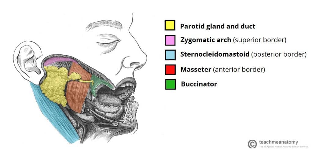
Fig 1.0 – Position of the parotid gland and borders of the parotid region.
Anatomical Relationships
The anatomical relationships of the parotid gland are of great clinical importance – particularly during parotid gland surgery.
Several important neurovascular structures pass through the gland:
- The facial nerve (cranial nerve VII), gives rise to five terminal branches within the parotid gland. These branches innervate the muscles of facial expression.
- The external carotid artery (ECA) ascends through the parotid gland. Within the gland, the ECA gives rise to the posterior auricular artery. The ECA then divides into its two terminal branches – the maxillary artery and superficial temporal artery.
- The retromandibular vein is formed within the parotid gland by the convergence of the superficial temporal and maxillary veins. It is one of the major structures responsible for venous drainage of the face.
By TeachMeSeries Ltd (2020)

Fig 1.1 – The branches of the facial nerve . The parotid gland has been removed to show their anatomical course.


